Spinolaminar Junction

Sonographically the pll is located approximately 4 6 mm deep anterior to the spinolaminar junction.
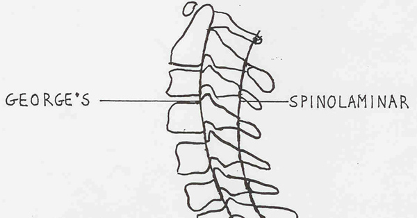
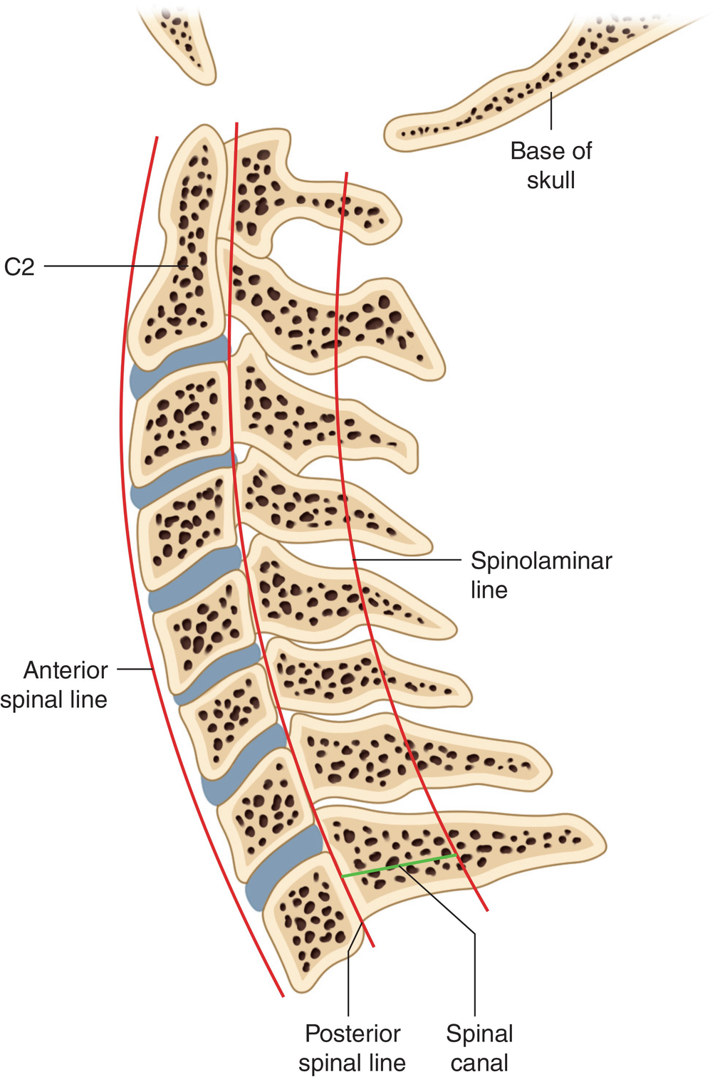
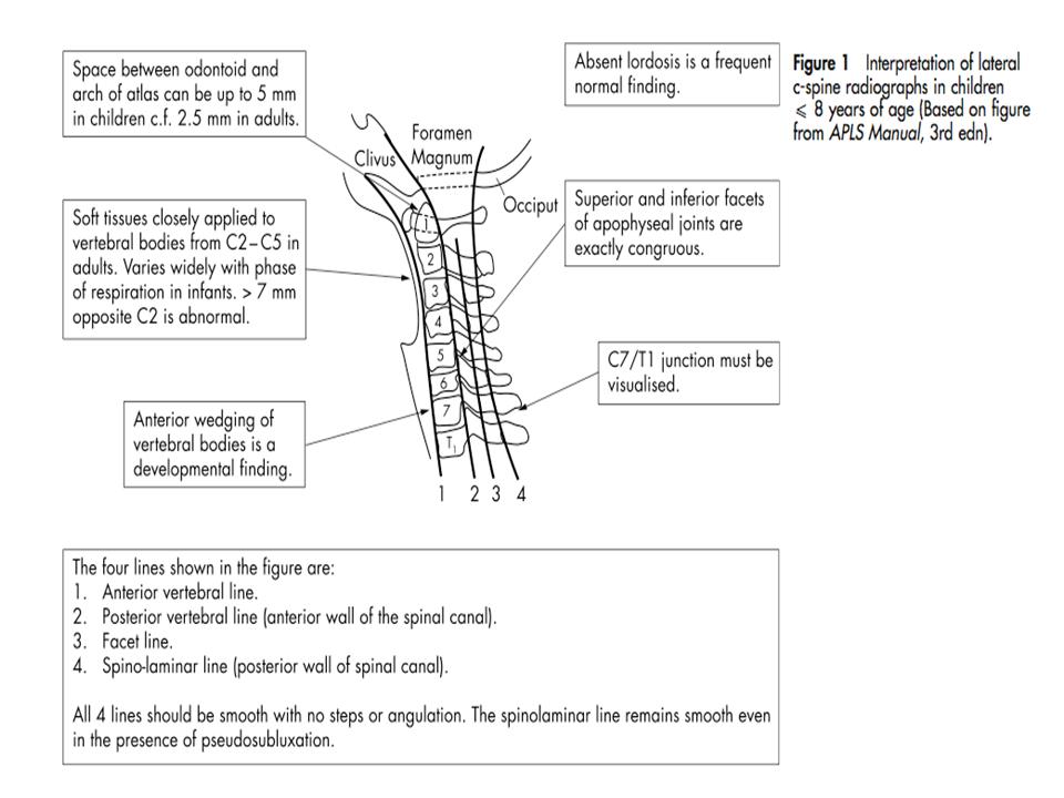
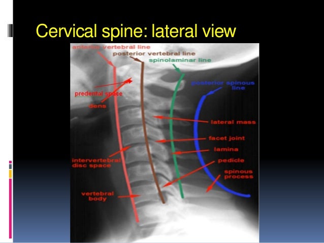
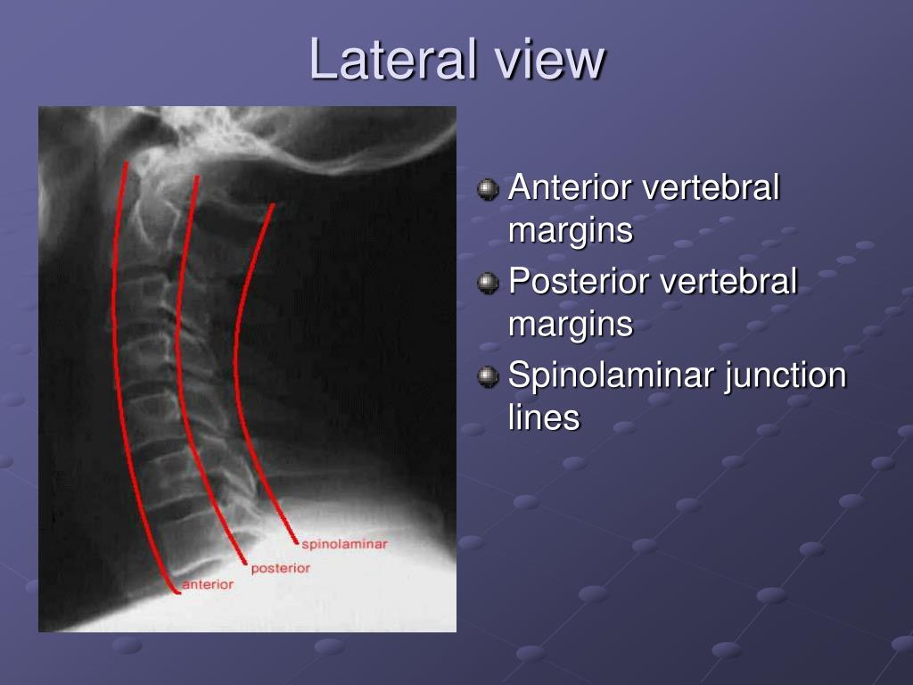
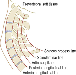
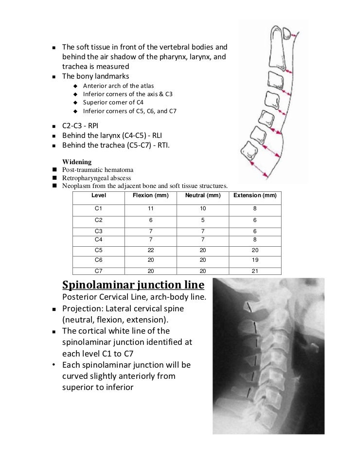
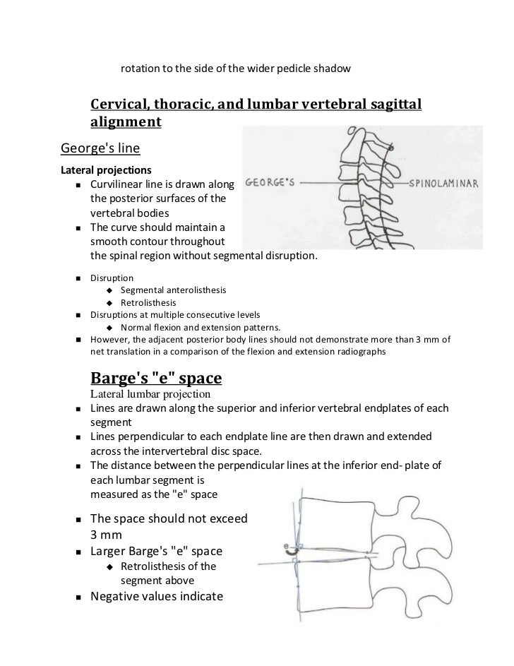
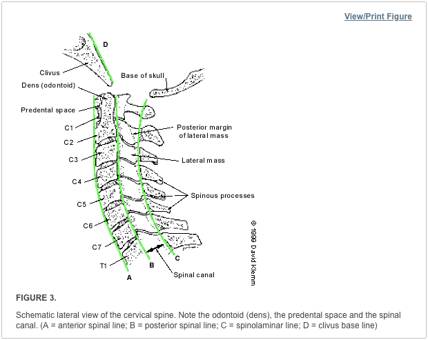
Spinolaminar junction. Posterior spinous line tips of the spinous processes these lines should follow a slightly lordotic curve smooth and without step offs. Spinolaminar line posterior margin of spinal canal 4. Any malalignment should be considered evidence of ligmentous injury or occult fracture and cervical spine immobilization should be maintained until a. A long high speed burr e g am 8 midas rex is then used to core out the cancellous and deep cortical surface of the contralateral lamina preserving the ligamentum flavum underneath and using it as a protective layer over the dura.
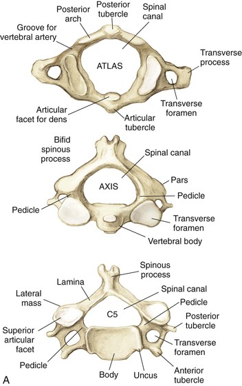
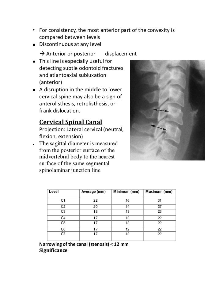
Formed by the junction of the c1 through c7 spinous processes and laminae marks the posterior boundary of the spinal canal. The distance from the posterior aspect of the ossicle to the spinolaminar junction line of c2 with the cervical spine in extension represented as dext is measured at 24 mm. The posterior arch of c1 may be a few millimeters anterior to the spinolaminar line as a normal variant. The reported union rate of the spinous process shows wide variations and there are conflicting opinions on whether it heals in a functional shape that provides a stable attachment site for the paravertebral muscles or merely fails to form a union at the spinolaminar junction resulting in a floated unattached spinous process with loss of the lever arm function as an attachment site for the paravertebral muscles.
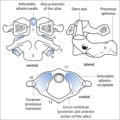
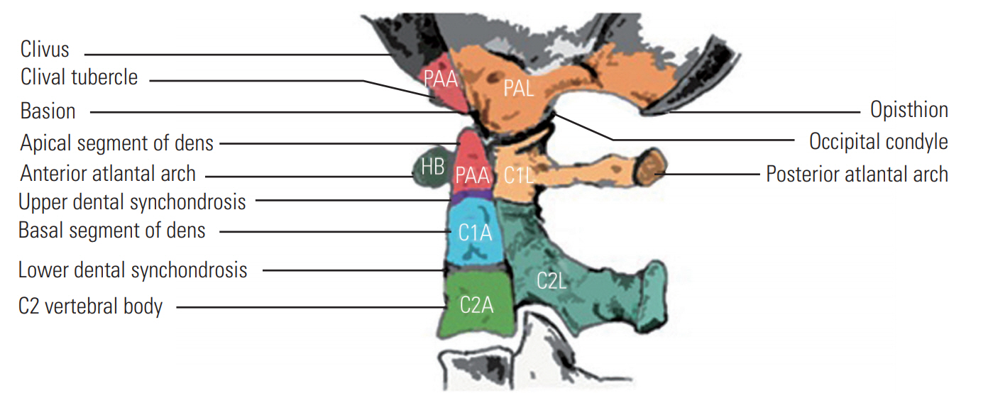
Midsagittal mdct image of the craniocervical junction demonstrates the powers ratio which is calculated by dividing the distance between the tip of the basion to the spinolaminar line by the distance from the tip of the opisthion to the midpoint of the posterior aspect of the anterior arch of c1. Spinolaminar line figure 6 6 c interspinous distances and posterior spinous processes figure 6 6 c midline craniocervical junction alignment figure 6 6 c basion dens interval c1 anterior arch dens interval opisthion posterior margin of foramen magnum c1 posterior arch c2 spinolaminar junction. Delineate the spinolaminar junction curve given a single reference point and the corners of each vertebrae. The line extends upward to the posterior rim of the foramen magnum.
Bovie cautery with a long tip is then used to remove the remaining muscle and soft tissue overlying the lamina and spinolaminar junction.

