Posterior Vitreous Detachment Ultrasound

Posterior Vitreous Detachment Jetem

Posterior Vitreous Detachment Jetem

Ultrasound Gel

Ultrasound Gel

Posterior Vitreous Detachment Radiology Reference Article Radiopaedia Org

Posterior Vitreous Detachment Radiology Reference Article Radiopaedia Org

Hennepin Ultrasound Vitreous Vs Retinal Detachment

Hennepin Ultrasound Vitreous Vs Retinal Detachment

Posterior Vitreous Detachment Radiology Reference Article Radiopaedia Org

Posterior Vitreous Detachment Radiology Reference Article Radiopaedia Org

Floaters Retinal Detachment Posterior Vitreous Detachment Or Vitreous Hemorrhage Emra

Floaters Retinal Detachment Posterior Vitreous Detachment Or Vitreous Hemorrhage Emra

Floaters Retinal Detachment Posterior Vitreous Detachment Or Vitreous Hemorrhage Emra

Posterior vitreous detachment pvd.

Posterior vitreous detachment ultrasound. If you see dark specks or flashes of light it s possible you could have posterior vitreous detachment pvd an eye problem many people have as they age. 8 the study has limitations but results suggest that with proper training ultrasound can be both sensitive and specific for identifying ocular pathology. One retrospective study of 130 pre operative ocular ultrasounds performed by a single operator reported 92 3 sensitivity and 98 3 specificity for identifying retinal detachment 96 2 and 100 for posterior vitreous detachment and 100 and 100 for vitreous hemorrhage. The condition is common for older adults.

Often high gain will allow abnormalities of the vitreous to be better visualized. To check ultrasound reliability in detecting retinal tears in pat. As you get older a gel inside your eye. Over 75 of those over the age of 65 develop it.

Ultrasound both a scan and contact b scan more useful ultrasound have a role in confirming the vitreous hemorrhage and also detecting underlying causes such as posterior vitreous detachment retinal detachment or tear trauma or malignancy 1 3. The vitreous body a clear gel like substance that fills the space between the lens and the retina microscopic fibers connect the vitreous body to the retina. Point of attachment referred to as the vitreous base. Vitreous hemorrhages vh and posterior vitreous detachments pvd will be apparent as the gain is increased.

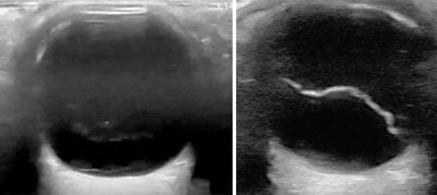
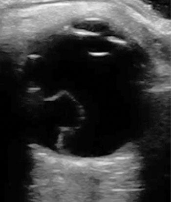
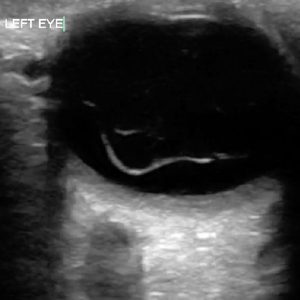
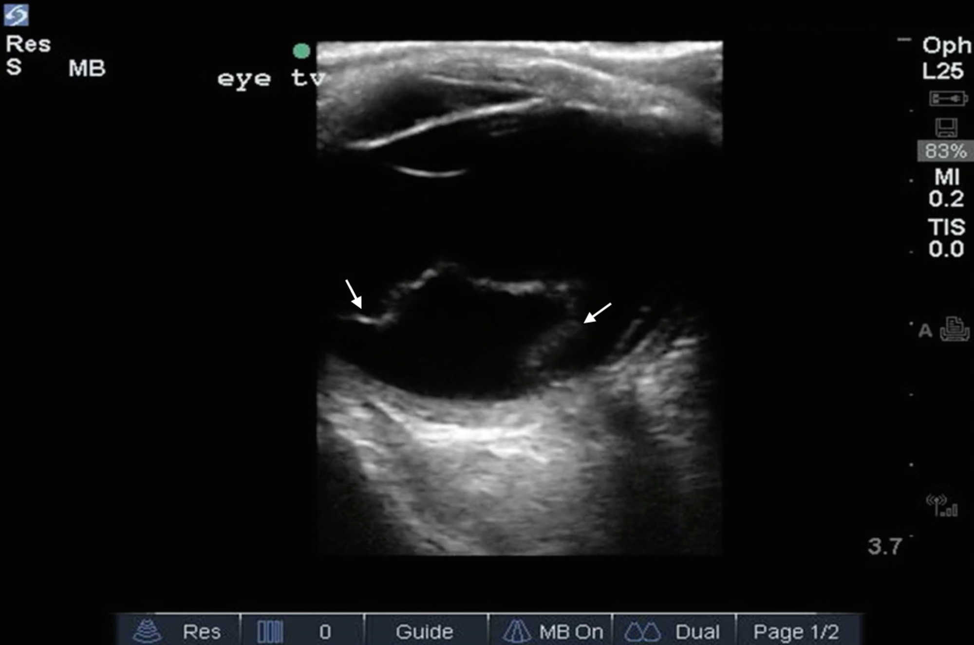
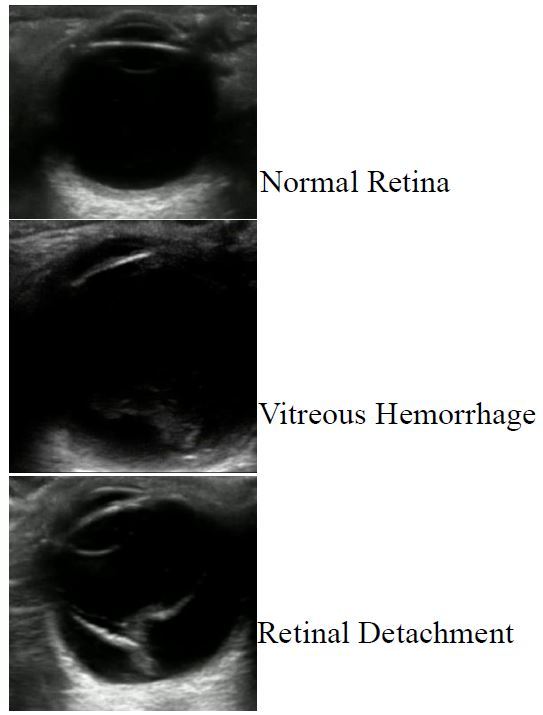
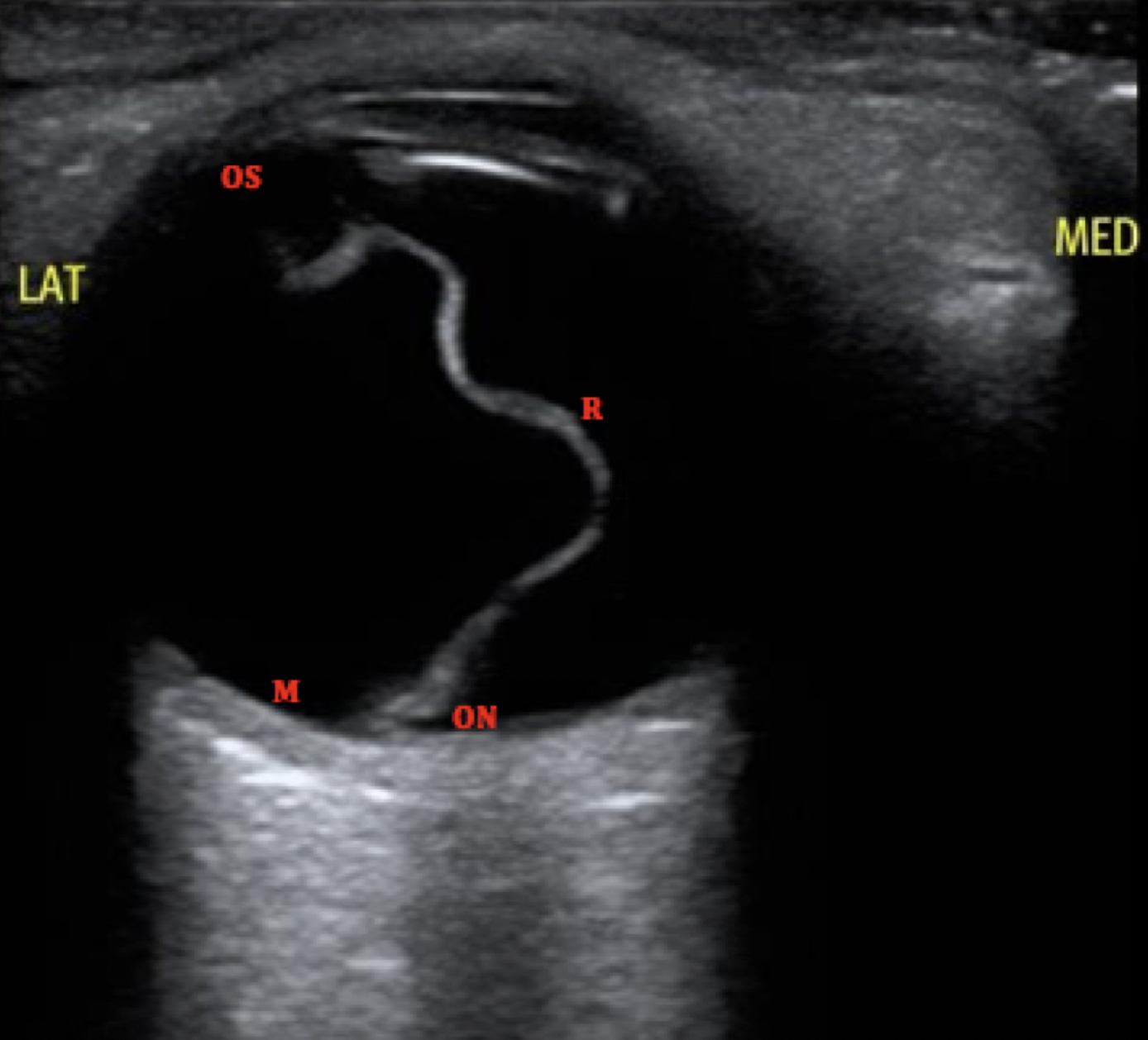
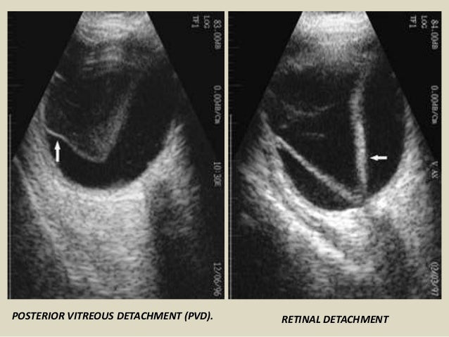
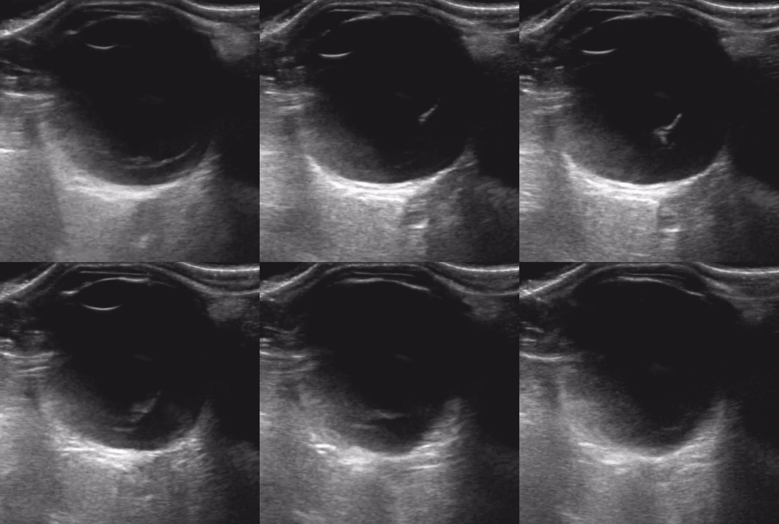
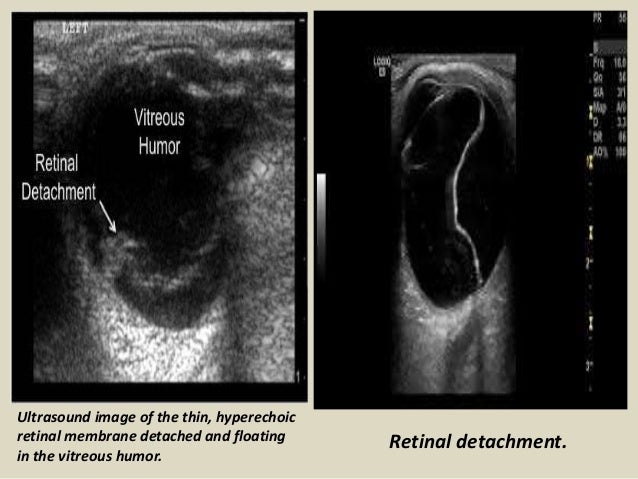
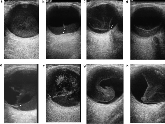
Linear echogenic membrane in the posterior compartment. Ultrasound can easily distinguish vitreous detachment from retinal detachment by looking to see if the detachment is tethered at the optic disc or not. In contrast to rd a pvd takes on a swaying seaweed appearance. The patient was discharged with no specific treatment.

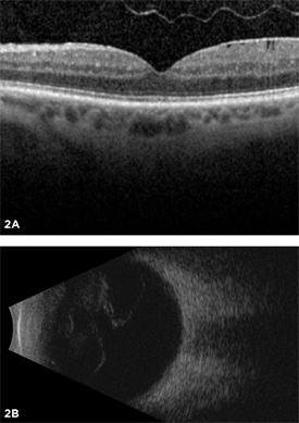
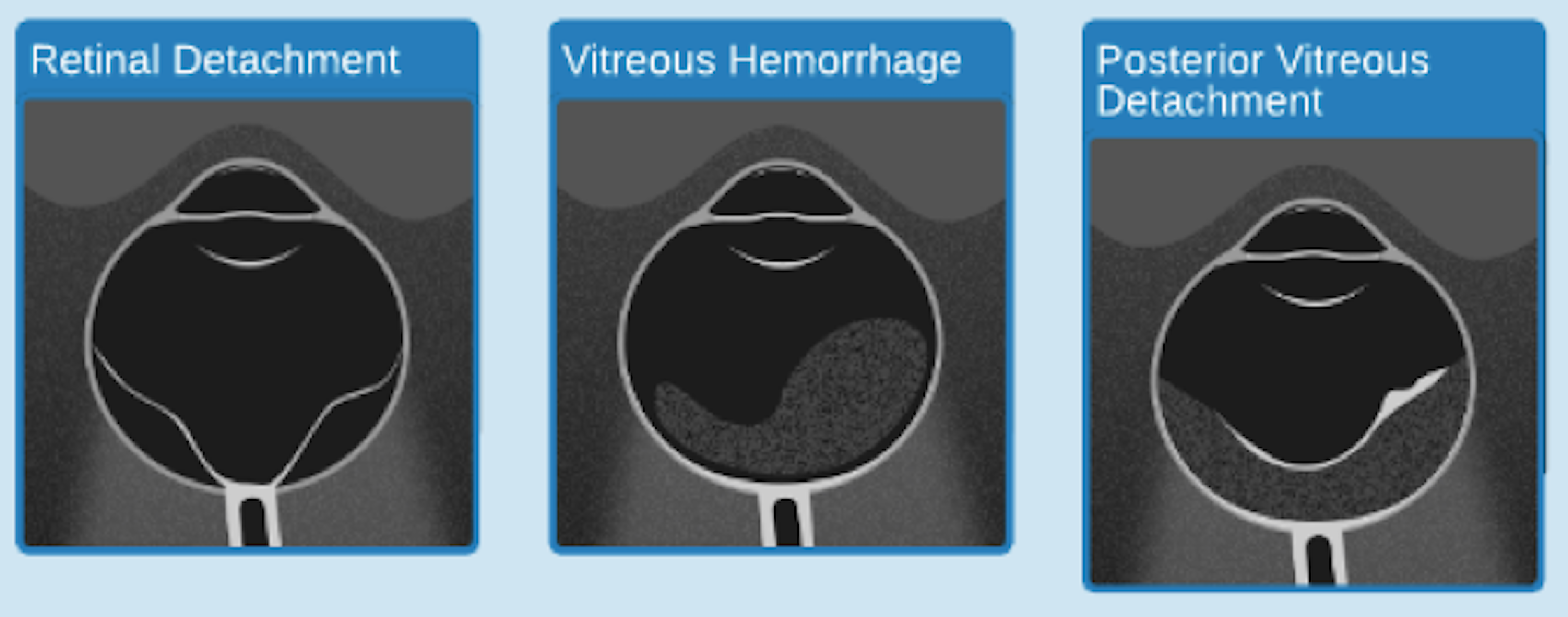
A posterior vitreous detachment is a condition of the eye in which the vitreous membrane separates from the retina. Although less common among people in their 40s or 50s the condition is not rare for those individuals. The sonographic appearance of isolated posterior vitreous detachment includes the following characteristics 3. Diagram of the vitreous cavity during posterior vitreous detachment.

Cross sectional study of 75 consecutive patients presenting with acute symptomatic posterior vitreous detachment aspvd and vitreous hemorrhage was conducted at university eye clinic university hospital sveti duh zagreb croatia. Once you have interrogated the vitreous body at normal settings slowly increase the gain. May demonstrate tethering near the ora serrata. It refers to the separation of the posterior hyaloid membrane from the retina anywhere posterior to the vitreous base.

Ophthalmology did see the patient in the ed and agreed that this was a posterior vitreous detachment.
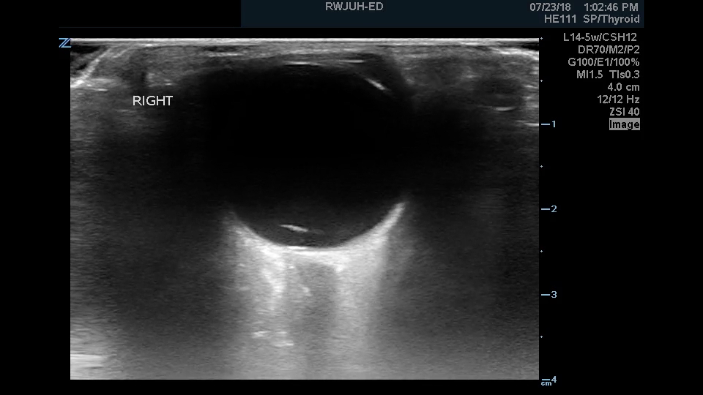
Ocular Ultrasound Posterior Vitreous Detachment Youtube

Ocular Ultrasound Posterior Vitreous Detachment Youtube

Ocular Ultrasound Retinal Detachment And Posterior Vitreous Detachment

Ocular Ultrasound Retinal Detachment And Posterior Vitreous Detachment

Ultrasound Example Of Posterior Vitreous Detachment Characterized By A Download Scientific Diagram

Ultrasound Example Of Posterior Vitreous Detachment Characterized By A Download Scientific Diagram

Floaters Retinal Detachment Posterior Vitreous Detachment Or Vitreous Hemorrhage Emra

Floaters Retinal Detachment Posterior Vitreous Detachment Or Vitreous Hemorrhage Emra

Retinal Detachment V Shaped Membrane Attached To Optic Disc Vitreous Hemorrhage Better To Be Assess Ultrasound Medical Ultrasound Vitreous Hemorrhage

Retinal Detachment V Shaped Membrane Attached To Optic Disc Vitreous Hemorrhage Better To Be Assess Ultrasound Medical Ultrasound Vitreous Hemorrhage

Figure 2 Vitreous Detachment May Appear Similar To A Retinal Detachment But Is More Globular And Not Tethered By Any Specific Structural Attachments Acep Now

Figure 2 Vitreous Detachment May Appear Similar To A Retinal Detachment But Is More Globular And Not Tethered By Any Specific Structural Attachments Acep Now

Cureus Non Traumatic Retinal Detachment Diagnosed Via Bedside Ultrasonography

Cureus Non Traumatic Retinal Detachment Diagnosed Via Bedside Ultrasonography

Bedside Ultrasound Of Retinal Detachment In A 19 Year Old Jetem

Bedside Ultrasound Of Retinal Detachment In A 19 Year Old Jetem

Eus Retinal Detachment Sstis Brown Emergency Medicine

Eus Retinal Detachment Sstis Brown Emergency Medicine

4 Posterior Vitreous Detachment Pvd In Two Patients A B Download Scientific Diagram

4 Posterior Vitreous Detachment Pvd In Two Patients A B Download Scientific Diagram

Retrospective Review Of Ocular Point Of Care Ultrasound For Detection Of Retinal Detachment The Western Journal Of Emergency Medicine

Retrospective Review Of Ocular Point Of Care Ultrasound For Detection Of Retinal Detachment The Western Journal Of Emergency Medicine

Posterior Vitreous Detachment The American Society Of Retina Specialists

Posterior Vitreous Detachment The American Society Of Retina Specialists

Ultrasound Gel

Ultrasound Gel

Https Encrypted Tbn0 Gstatic Com Images Q Tbn 3aand9gcrt0fgnc K8ni2gxn R1v773gtio Vxeuws3a Usqp Cau

Https Encrypted Tbn0 Gstatic Com Images Q Tbn 3aand9gcrt0fgnc K8ni2gxn R1v773gtio Vxeuws3a Usqp Cau

Macula Off Retinal Detachment Identified On Bedside Ultrasound Jetem

Macula Off Retinal Detachment Identified On Bedside Ultrasound Jetem

A High Resolution 7 5 10 Mhz Or Higher Linear Array Ultrasound Transducer Is Used

A High Resolution 7 5 10 Mhz Or Higher Linear Array Ultrasound Transducer Is Used

Ocular Ultrasound Core Em

Ocular Ultrasound Core Em

Echography Ultrasound Eyewiki

Echography Ultrasound Eyewiki

1

1

Floaters Retinal Detachment Posterior Vitreous Detachment Or Vitreous Hemorrhage Emra

Floaters Retinal Detachment Posterior Vitreous Detachment Or Vitreous Hemorrhage Emra

Presentation1 Pptx Ultrasound Examination Of The Orbit

Presentation1 Pptx Ultrasound Examination Of The Orbit

Ocular Ultrasound Retinal Detachment And Posterior Vitreous Detachment

Ocular Ultrasound Retinal Detachment And Posterior Vitreous Detachment

Https Journals Sagepub Com Doi Pdf 10 1177 1024907917735085

Https Journals Sagepub Com Doi Pdf 10 1177 1024907917735085

Https Encrypted Tbn0 Gstatic Com Images Q Tbn 3aand9gcq6k Bwjeaeowox5tpeg0 Ixovw0hha C6qda Usqp Cau

Https Encrypted Tbn0 Gstatic Com Images Q Tbn 3aand9gcq6k Bwjeaeowox5tpeg0 Ixovw0hha C6qda Usqp Cau

Presentation1 Pptx Ultrasound Examination Of The Orbit

Presentation1 Pptx Ultrasound Examination Of The Orbit

Vitreous Retina Adhesion In A Case Of Posterior Vitreous Detachment Download Scientific Diagram

Vitreous Retina Adhesion In A Case Of Posterior Vitreous Detachment Download Scientific Diagram

Imaging The Vitreous Optical Coherence Tomography And Ultrasound Imaging Eye

Imaging The Vitreous Optical Coherence Tomography And Ultrasound Imaging Eye

Https Encrypted Tbn0 Gstatic Com Images Q Tbn 3aand9gcsm4dspx7udsdqxu1crvfro4t4pzp6rc6sc6a Usqp Cau

Https Encrypted Tbn0 Gstatic Com Images Q Tbn 3aand9gcsm4dspx7udsdqxu1crvfro4t4pzp6rc6sc6a Usqp Cau

Ultrasound Of Retinal Vs Vitreous Detachment Youtube

Ultrasound Of Retinal Vs Vitreous Detachment Youtube

Accuracy Of Spectral Domain Oct Of The Macula For Detection Of Complete Posterior Vitreous Detachment Ophthalmology Retina

Accuracy Of Spectral Domain Oct Of The Macula For Detection Of Complete Posterior Vitreous Detachment Ophthalmology Retina

A Ocular Ultrasound Of A Normal Eye Is Shown B A Retinal Flap Download Scientific Diagram

A Ocular Ultrasound Of A Normal Eye Is Shown B A Retinal Flap Download Scientific Diagram

Echography Ultrasound Eyewiki

Echography Ultrasound Eyewiki

Djo Digital Journal Of Ophthalmology

Djo Digital Journal Of Ophthalmology

Https Retinatoday Com Pdfs 1109 08 Pdf

Https Retinatoday Com Pdfs 1109 08 Pdf

Source : pinterest.com