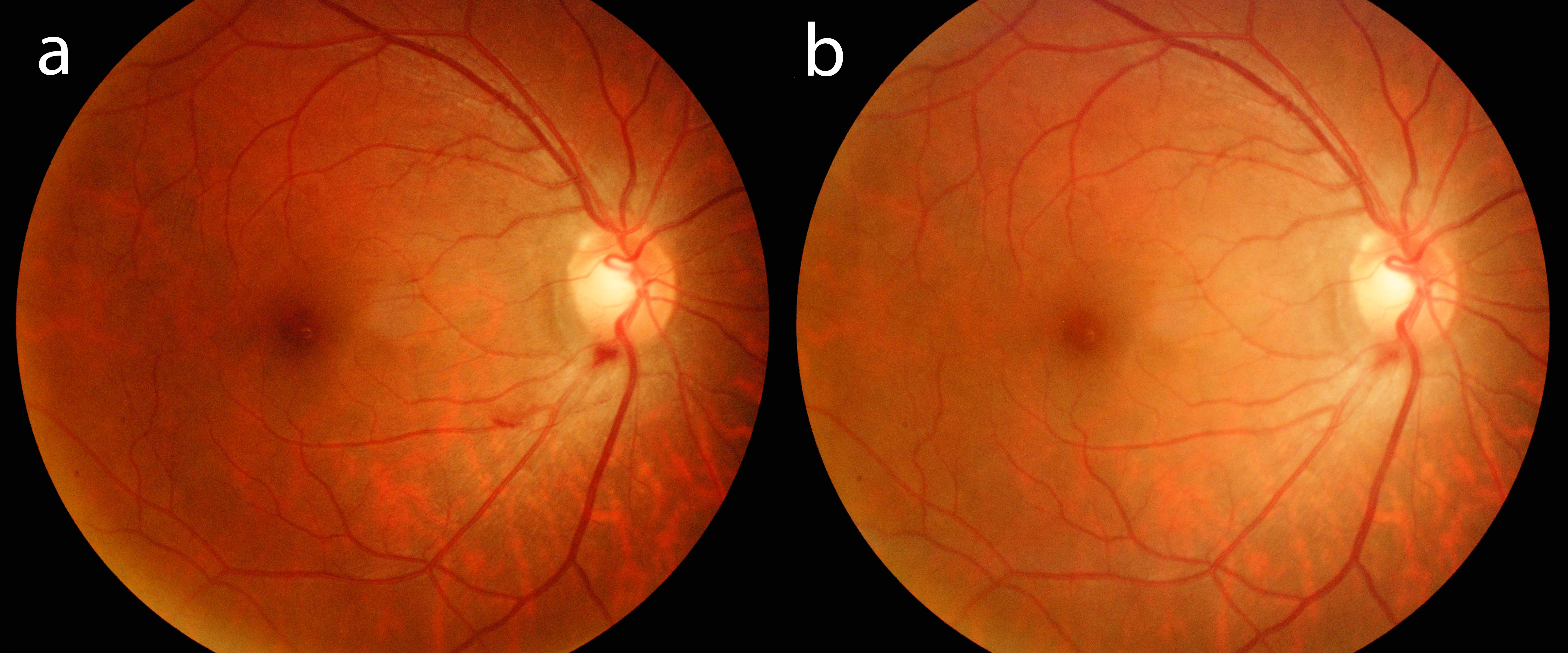
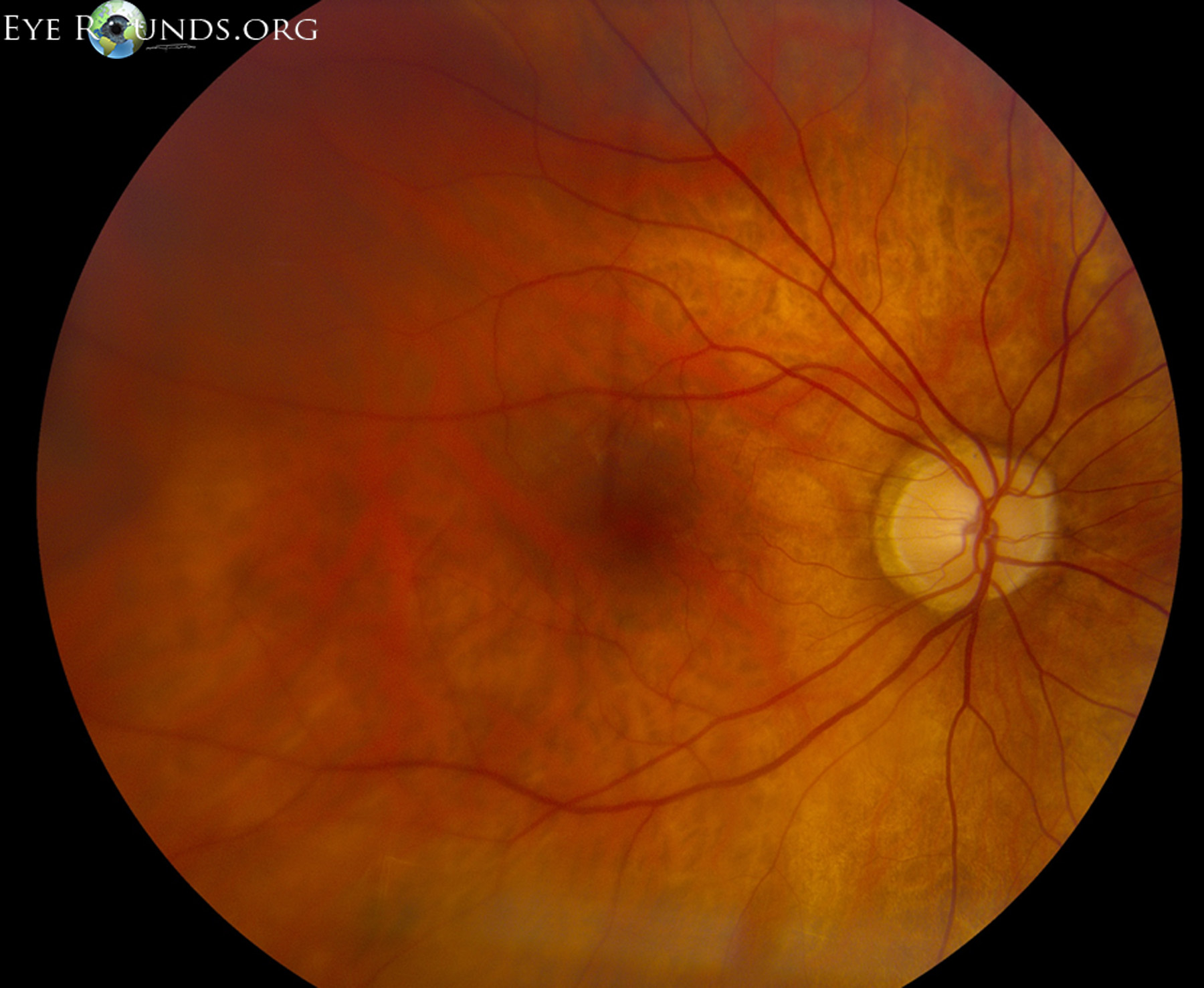
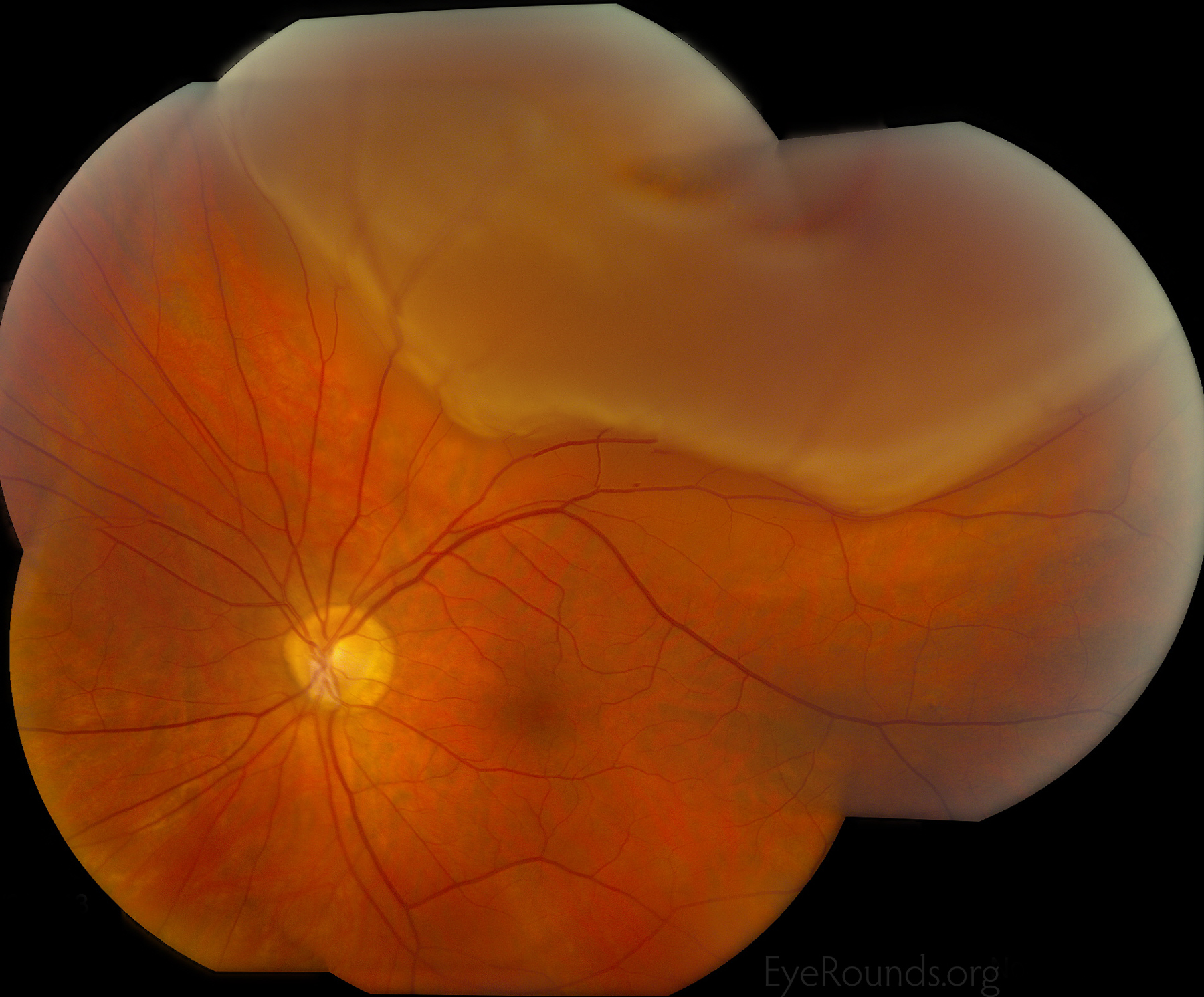
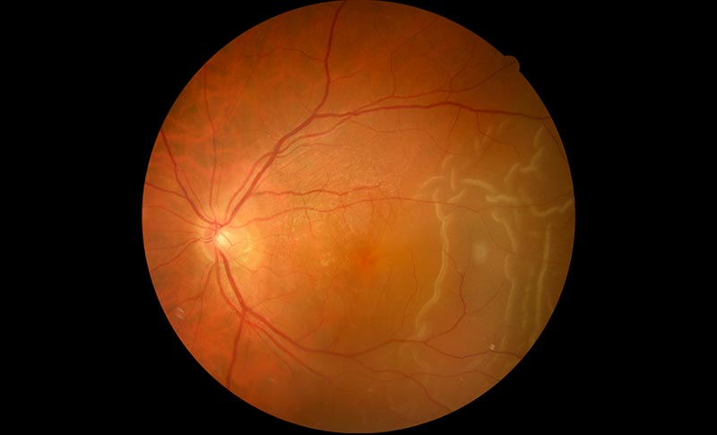
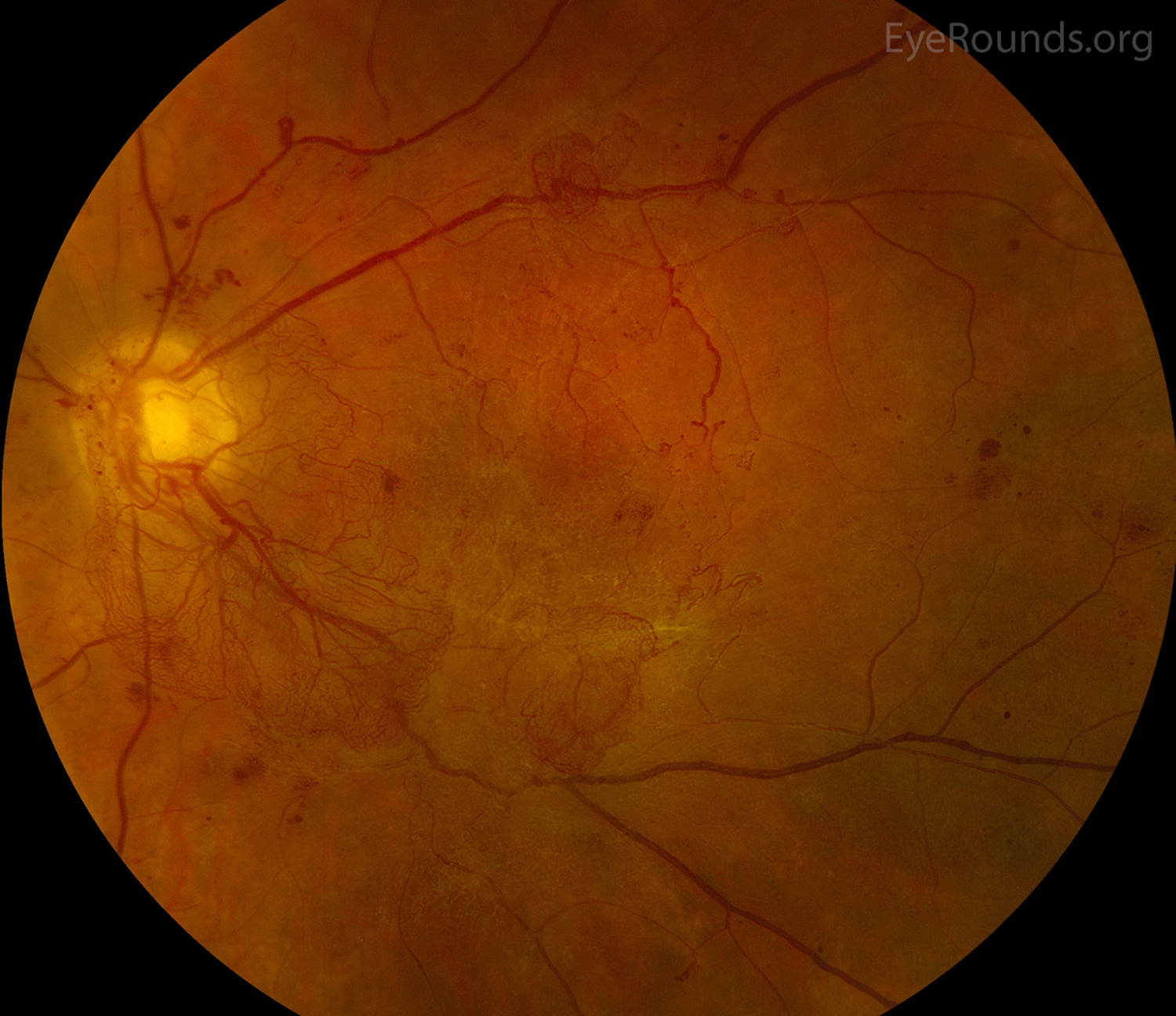
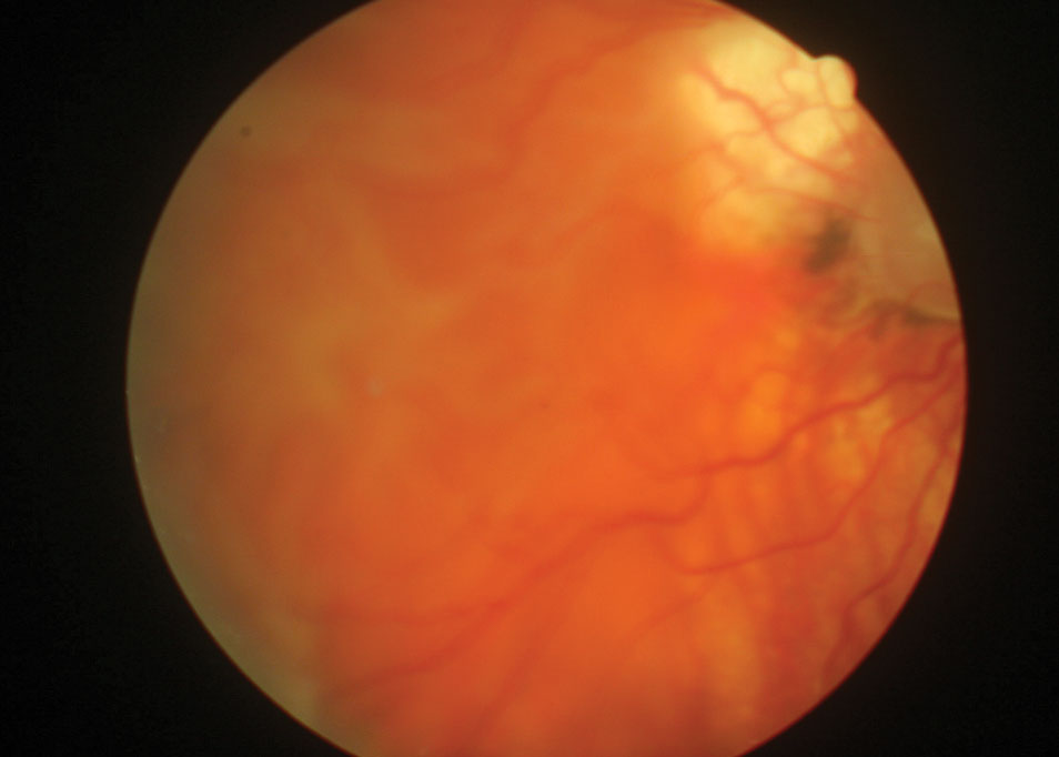
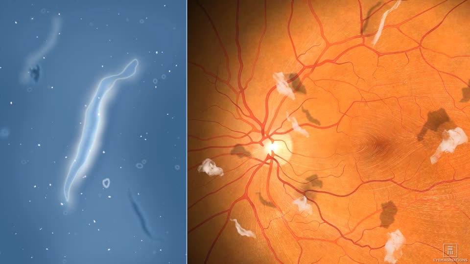
Posterior Vitreous Detachment Fundus

Vitreous Syneresis An Impending Posterior Vitreous Detachment Pvd

Vitreous Syneresis An Impending Posterior Vitreous Detachment Pvd

Vitreous Syneresis An Impending Posterior Vitreous Detachment Pvd

Vitreous Syneresis An Impending Posterior Vitreous Detachment Pvd

Cureus Posterior Vitreous Detachment Precipitated By Yoga

Cureus Posterior Vitreous Detachment Precipitated By Yoga

Vitreous Syneresis An Impending Posterior Vitreous Detachment Pvd

Vitreous Syneresis An Impending Posterior Vitreous Detachment Pvd

Vitreous Syneresis An Impending Posterior Vitreous Detachment Pvd

Vitreous Syneresis An Impending Posterior Vitreous Detachment Pvd

Evaluation And Management Of Pvd

Evaluation And Management Of Pvd

Evaluation And Management Of Pvd

Therefore it is important to be examined shortly after these symptoms begin.

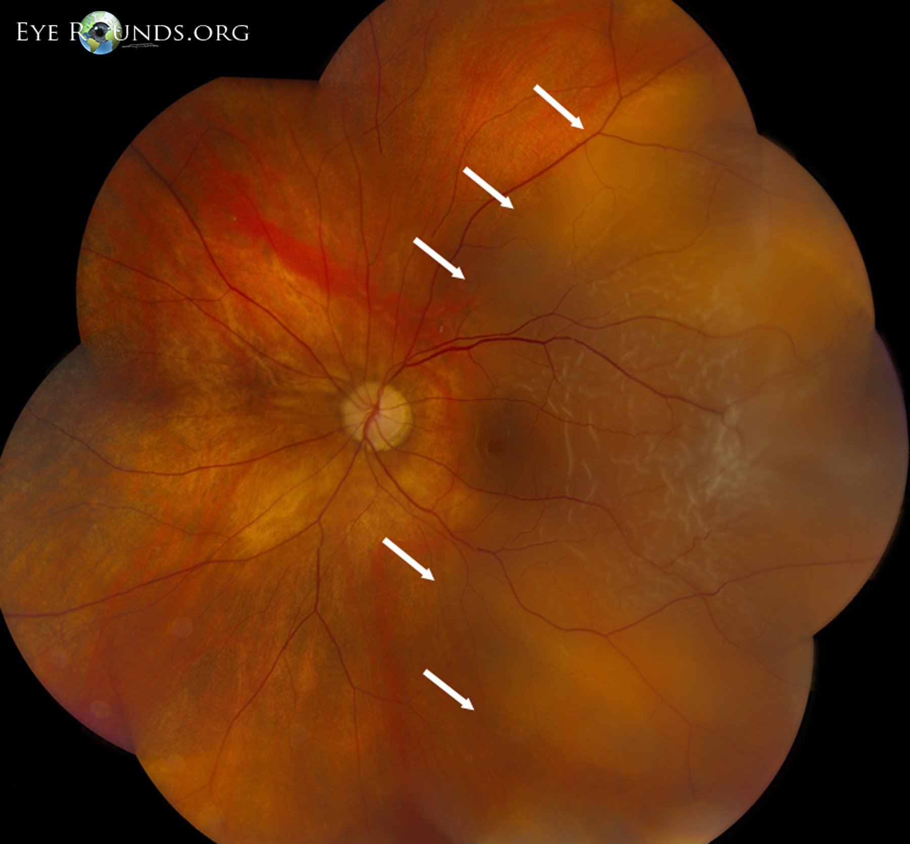
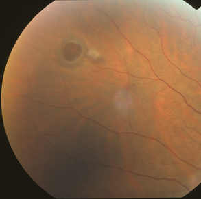
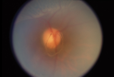
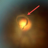
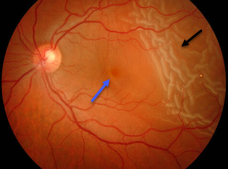
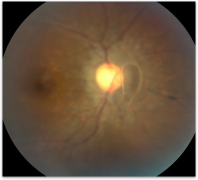
Posterior vitreous detachment fundus. If you see dark specks or flashes of light it s possible you could have posterior vitreous detachment pvd an eye problem many people have as they age. Microscopic fibers connect the vitreous body to the retina. Posterior vitreous detachment pvd is a separation between the posterior vitreous cortex and the neurosensory retina with the vitreous collapsing anteriorly towards the vitreous base. In most cases a vitreous detachment also known as a posterior vitreous detachment is not sight threatening and requires no treatment.

Some research has found that the condition is more common among women. Pvd is common and occurs naturally. If a retinal tear does occur during a posterior vitreous detachment it usually happens at the same time as one begins to experience symptoms of the pvd. Usually the fibers break allowing the vitreous to separate and shrink from the retina.

And sounds a bit scary fortunately this eye condition usually won t threaten your vision or require treatment. It refers to the separation of the posterior hyaloid membrane from the retina anywhere posterior to the vitreous base. Posterior vitreous detachment is quite a mouthful. This is a vitreous detachment.

Posterior vitreous detachment pvd occurs when the vitreous shrinks and pulls away from the retina. Although less common among people in their 40s or 50s the condition is not rare for those individuals. A posterior vitreous detachment is a condition of the eye in which the vitreous membrane separates from the retina. Download fact sheet large print version spanish translation.

The condition is common for older adults. But it can sometimes signal a more serious sight threatening problem. Posterior vitreous detachment pvd is a natural change that occurs during adulthood when the vitreous gel that fills the eye separates from the retina the light sensing nerve layer at the back of the eye. This can cause floaters and flashes to appear more frequently in your vision.

Posterior vitreous detachment pvd occurs when the fibers in your eye s vitreous layer shrink and condense causing the vitreous gel to pull on the retina s surface. A vitreous detachment is a common condition that usually affects people over age 50 as the vitreous shrinks it becomes somewhat stringy and the strands can cast tiny shadows on the retina that you may notice as floaters which appear as little cobwebs or specks that seem to float about in your field of vision. As you get older a gel inside your eye.
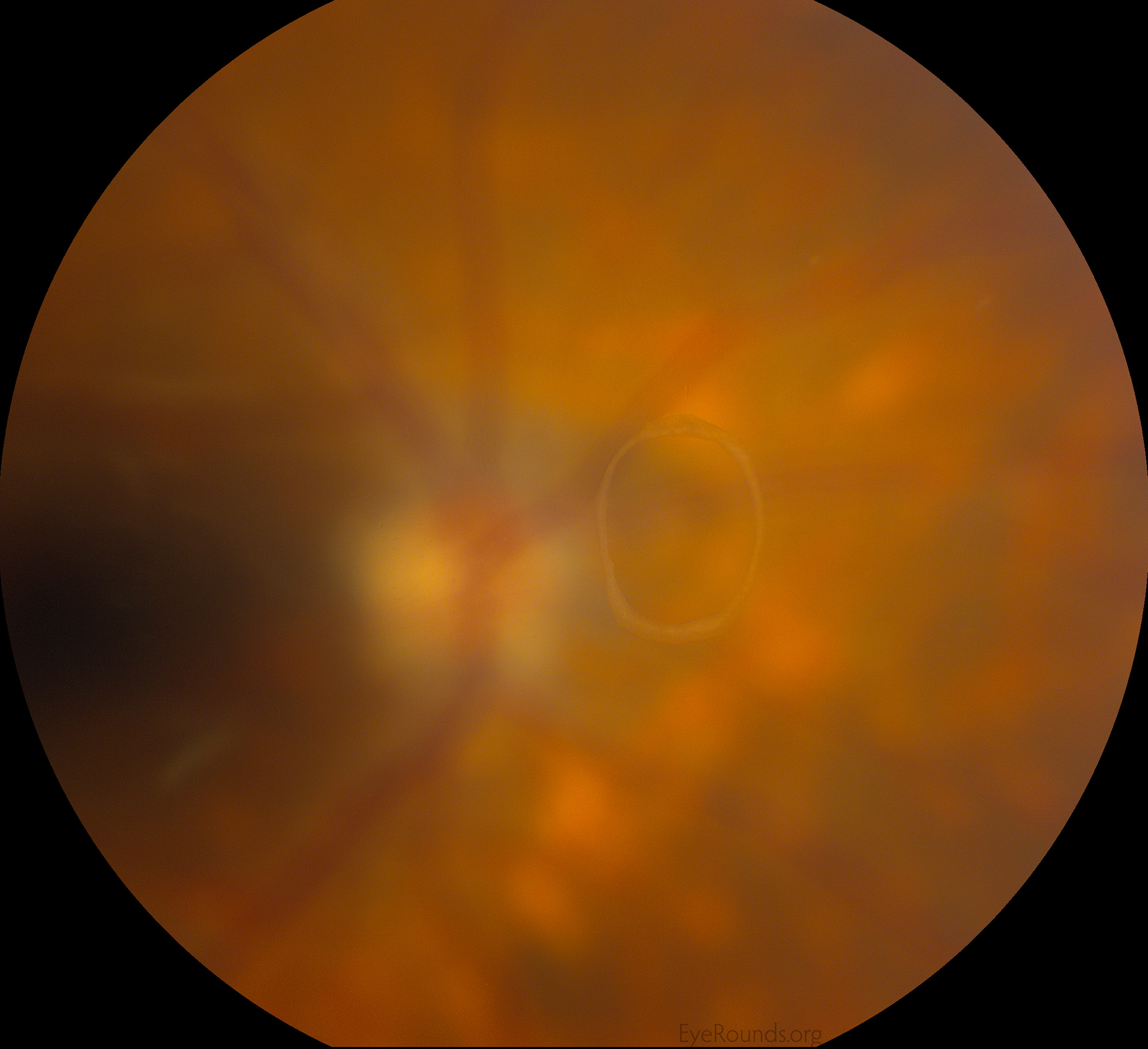
Vitreous Syneresis An Impending Posterior Vitreous Detachment Pvd

Vitreous Syneresis An Impending Posterior Vitreous Detachment Pvd

Evaluation And Management Of Pvd

Evaluation And Management Of Pvd

Peripapillary Hemorrhage Associated With Posterior Vitreous Detachment The Retina Reference

Peripapillary Hemorrhage Associated With Posterior Vitreous Detachment The Retina Reference

Posterior Vitreous Detachment Retina Image Bank

Posterior Vitreous Detachment Retina Image Bank

Posterior Vitreous Detachment Wikipedia

Posterior Vitreous Detachment Wikipedia

Posterior Vitreous Detachment Posterior Vitreous Detachment Eye Health Eye Doctor

Posterior Vitreous Detachment Posterior Vitreous Detachment Eye Health Eye Doctor

Retina Eye Specialists Www Retinaeye Com

Retina Eye Specialists Www Retinaeye Com

Optometric Management Clinical Retina

Optometric Management Clinical Retina

Evaluation And Management Of Pvd

Evaluation And Management Of Pvd

Retina Louisville Posterior Vitreous Detachment Bennett Bloom

Retina Louisville Posterior Vitreous Detachment Bennett Bloom

Diagnosis And Treatment Of Retinal Detachment In Gironde

Diagnosis And Treatment Of Retinal Detachment In Gironde

Posterior Vitreous Detachment Retina Image Bank

Posterior Vitreous Detachment Retina Image Bank

Variations Of Posterior Vitreous Detachment British Journal Of Ophthalmology

Variations Of Posterior Vitreous Detachment British Journal Of Ophthalmology

Vitreous Hemorrhage From One Medical Student To Another

Vitreous Hemorrhage From One Medical Student To Another

Fundus Photograph Of Pvd The Detached Posterior Vitreous Cortex Download Scientific Diagram

Fundus Photograph Of Pvd The Detached Posterior Vitreous Cortex Download Scientific Diagram

Racgp Flashes And Floaters A Practical Approach To Assessment And Management

Racgp Flashes And Floaters A Practical Approach To Assessment And Management

Retinal Detachment Baton Rouge La Ophthalmologist

Retinal Detachment Baton Rouge La Ophthalmologist

That S A Foul

That S A Foul

Https Encrypted Tbn0 Gstatic Com Images Q Tbn 3aand9gcrrairuikii6sxeput 8w48juawxmlfmxk Ph1avkaaap49d Jp Usqp Cau

Https Encrypted Tbn0 Gstatic Com Images Q Tbn 3aand9gcrrairuikii6sxeput 8w48juawxmlfmxk Ph1avkaaap49d Jp Usqp Cau

Diagnosis And Treatment Of Posterior Vitreous Detachment

Diagnosis And Treatment Of Posterior Vitreous Detachment

Primary Retinal Detachment Nejm

Primary Retinal Detachment Nejm

Posterior Vitreous Detachment Ento Key

Posterior Vitreous Detachment Ento Key

Racgp Flashes And Floaters A Practical Approach To Assessment And Management

Racgp Flashes And Floaters A Practical Approach To Assessment And Management

Retina Louisville Retinal Detachment Louisville Bennett Bloom

Retina Louisville Retinal Detachment Louisville Bennett Bloom

Posterior Vitreous Detachment Retina Vitreous Surgeons Of Cny

Posterior Vitreous Detachment Retina Vitreous Surgeons Of Cny

Weiss Ring Retina Image Bank

Weiss Ring Retina Image Bank

Primary Rhegmatogenous Retinal Detachment Ento Key

Primary Rhegmatogenous Retinal Detachment Ento Key

Full Text Classification Of Posterior Vitreous Detachment Opth

Full Text Classification Of Posterior Vitreous Detachment Opth

Pvd Confirms The Worst

Pvd Confirms The Worst

Fundus Image Shows Induction Of Posterior Vitreous Detachment Download Scientific Diagram

Fundus Image Shows Induction Of Posterior Vitreous Detachment Download Scientific Diagram

Moran Core Red Flag Symptoms Flashes Floaters Curtains

Moran Core Red Flag Symptoms Flashes Floaters Curtains

Vitreous Floaters Eyeonoptics

Vitreous Floaters Eyeonoptics

Common Eye Emergencies American Family Physician

Common Eye Emergencies American Family Physician

Malpractice Risk Retinal Detachments American Academy Of Ophthalmology

Malpractice Risk Retinal Detachments American Academy Of Ophthalmology

Source : pinterest.com